
Introduction
Gestational trophoblastic disease (GTD) encompasses a spectrum of placental trophoblastic disorders. While complete hydatidiform mole (CHM) classically presents with vaginal bleeding or hyperemesis, early diagnosis can be challenging when the patient is completely asymptomatic, presenting instead as a pregnancy of an unknown location (PUL). The primary novelty of this case lies in the complete absence of clinical symptoms in an adolescent patient whose initial non-diagnostic PUL ultrasound rapidly evolved into classic molar sonographic architecture on repeat evaluation.
Case Presentation
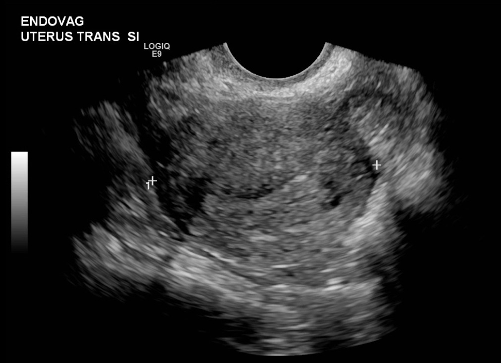
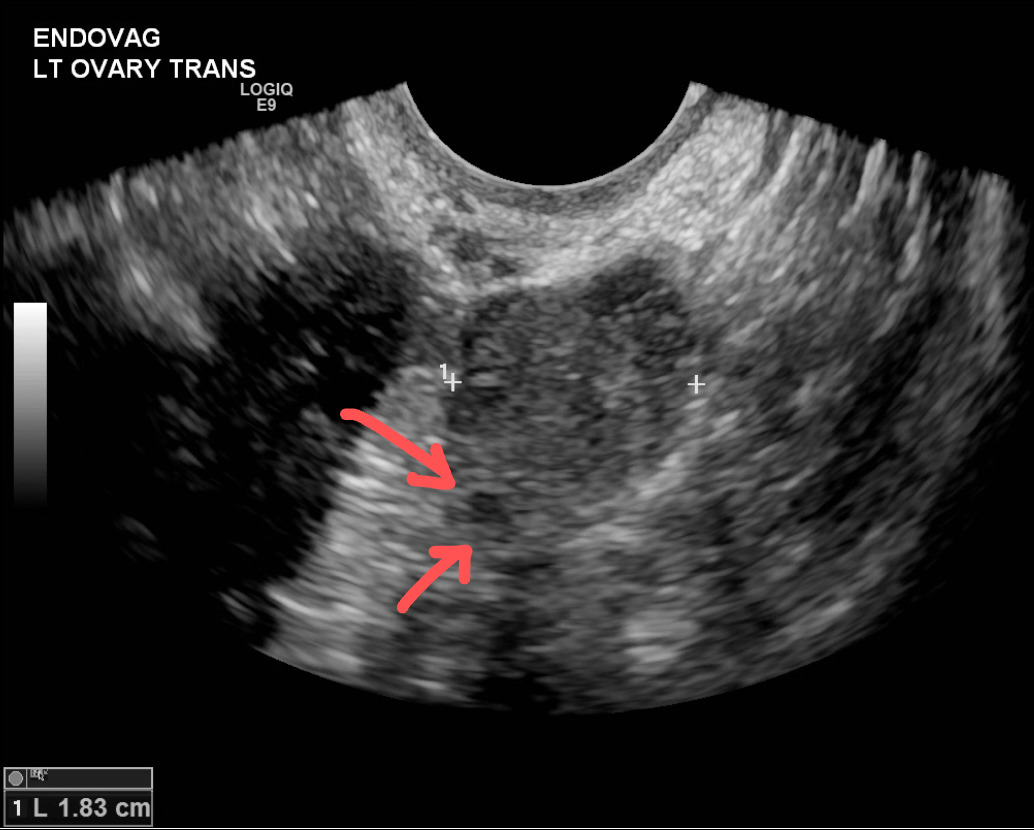
A 17-year-old G1P0 female was referred to the Emergency Department at an estimated 7 weeks’ gestation, based on last menstrual period, following a non-diagnostic outpatient ultrasound. She was entirely asymptomatic, reporting no vaginal bleeding, abdominal pain, or hyperemesis. On arrival, vital signs were stable (BP 118/78 mmHg, HR 72 bpm, RR 16bpm, T 99F, SpO2 99% on room air) with an unremarkable abdominal examination. Pelvic examination was deferred in the absence of vaginal bleeding or pelvic discomfort. Initial workup revealed a quantitative β-hCG level of 13,179 mIU/mL. Formal pelvic ultrasound found the uterus measured 6.6×3.6×4.6cm and was inconclusive for intrauterine pregnancy; a 1.8cm left corpus luteal cyst was noted (Figures 1A and 1B). Given her clinical stability and indeterminate imaging findings, the OB/GYN service was consulted, with a recommendation for discharge with strict return precautions and a return visit within 48-72 hours for repeat β-hCG and imaging.
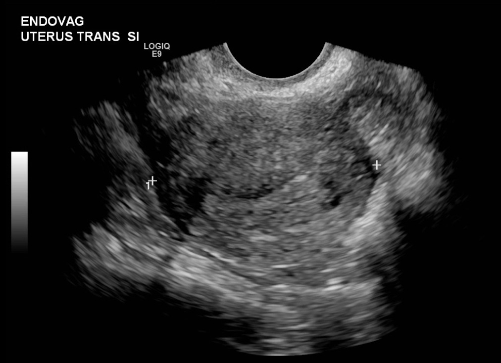
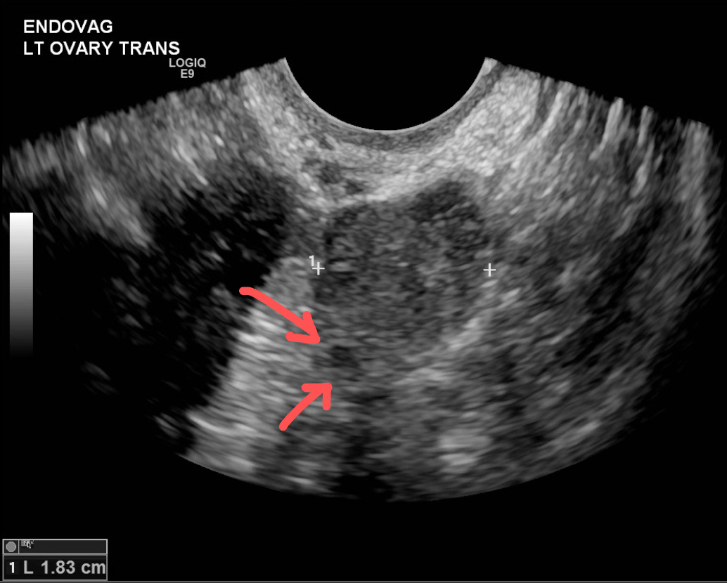
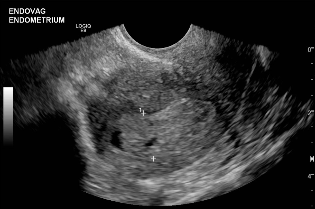
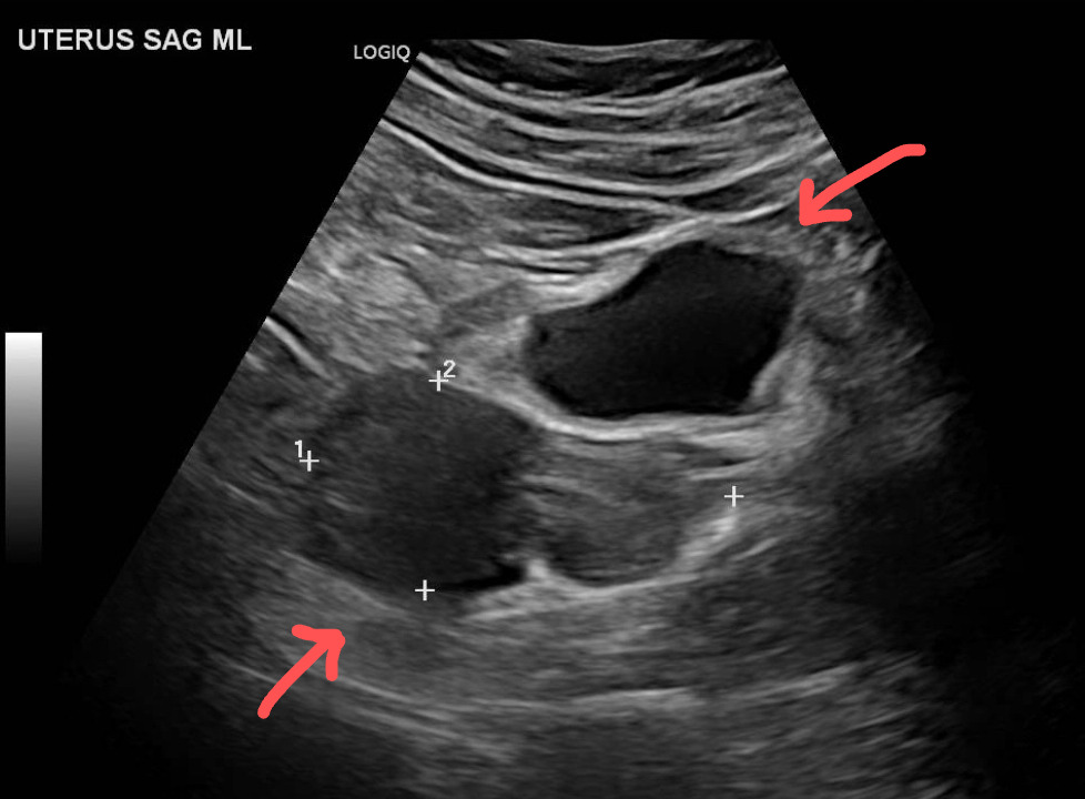
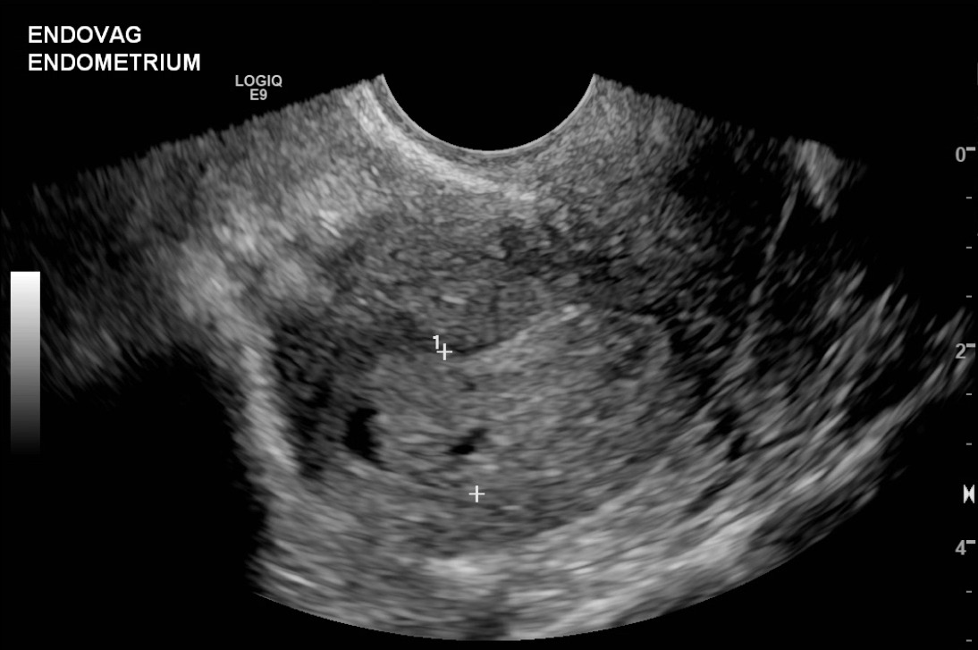
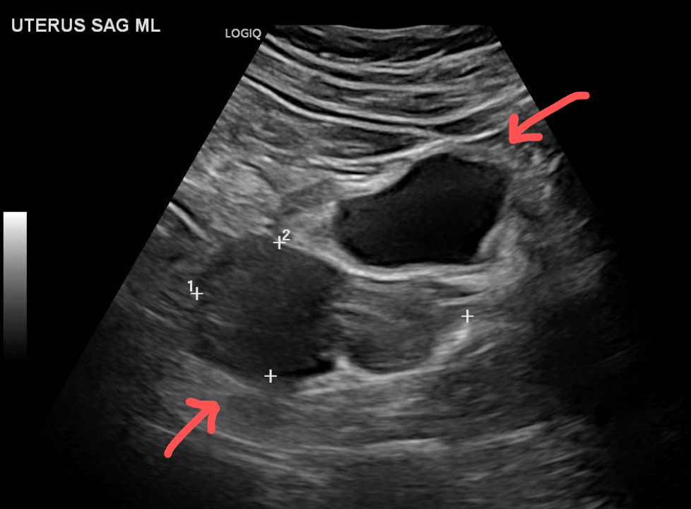
The patient returned 72 hours later, remaining completely asymptomatic with no new complaints. Her vital signs and physical examination were unchanged from the initial visit (BP 115/75 mmHg, HR 70 bpm, RR 15 bpm, T 98.1 F, SpO2 100% on room air), and her abdominal examination remained benign; pelvic examination was again deferred due to lack of vaginal bleeding and pelvic discomfort. Repeat laboratory analysis revealed a quantitative β-hCG level of 19,977 mIU/mL, representing a 51.6% rise, suboptimal for a normal viable intrauterine pregnancy, yet non-declining like a spontaneous abortion. Ancillary labs (CBC, CMP, and urinalysis) remained unremarkable, reinforcing clinical stability, but not excluding a pathological process. Repeat formal TVUS revealed a uterus measuring 8.1×4.0×5.8cm (Figure 2A) containing a markedly thickened, heterogeneous endometrial mass with microcystic spaces, producing the classic vesicular “snowstorm” appearance (Figure 2B). Spectral and color Doppler evaluation demonstrated minimal vascularity within the microcystic spaces. No extrauterine gestation or pelvic free fluid was appreciated. The combination of the sub-optimal β-hCG rise, increased uterine size, and characteristic ultrasound findings prompted the presumptive diagnosis of gestational trophoblastic disease and urgent Gynecologic-oncology consultation.
Gynecology Oncology was consulted and confirmed that she was clinically stable, allowing for outpatient intervention. The patient was discharged and later returned for outpatient suction dilation and curettage (D&C) without complication (estimated blood loss of 150mL). Histopathology confirmed the definitive diagnosis as a Complete Hydatidiform Mole. The patient was discharged with a mandatory follow-up for serial β-hCG surveillance for complete resolution and to guide management aimed at preventing the development of Gestational Trophoblastic Neoplasia (GTN). The patient was compliant and had an undetectable β-hCG level several weeks later.
Discussion
An asymptomatic pregnancy of an unknown location (PUL) with elevated β-hCG represents a diagnostic challenge in the emergency department.1 Standard evaluation requires systematic differentiation among four primary clinical entities: early viable intrauterine pregnancy (expected β-hCG doubling), failed early pregnancy (appropriately falling β-hCG), ectopic pregnancy (plateauing β-hCG with extrauterine findings), and early gestational trophoblastic disease (non-doubling β-hCG with delayed intrauterine trophoblastic proliferation).2–4 Under established consensus guidelines, the discriminatory zone for transvaginal ultrasound visualization of an intrauterine gestational sac is generally 1,500-2,000 mIU/mL, with conservative thresholds extending up to 3,500 mIU/mL.1 In this patient, the initial β-hCG of 13,179 mIU/mL far exceeded the discriminatory threshold without a visible IUP, establishing a high-risk PUL that mandated close short-interval surveillance.
While classic, advanced complete hydatidiform mole (CHM) presents with β-hCG > 100,000 mIU/mL, early complete moles or those presenting as PUL frequently present during early logarithmic growth or plateau phases.1,5 In this patient, the 51.6% rise over 72 hours represented a suboptimal trajectory relative to normal intrauterine gestation, serving as the key diagnostic prompt to repeat formal imaging despite the complete lack of symptoms.1,6 Additionally, because extreme β-hCG elevations can cross-react with thyroid-stimulating hormone (TSH) receptors, obtaining baseline thyroid function tests in high β-hCG presentations is recommended to exclude occult thyrotoxicosis.7
Point-of-care ultrasound (POCUS) allows emergency clinicians to rapidly assess intra-abdominal hemorrhage or overt intrauterine pathology.6,8 When initial imaging is non-diagnostic in a clinically stable patient, structured outpatient surveillance with standardized return precautions is safe and effective.4 Once characteristic sonographic features appear, prompt specialty consultation ensures controlled, non-emergent surgical intervention.8,9
Conclusion
This case highlights that early complete hydatidiform mole can present silently as an asymptomatic pregnancy of unknown location with a suboptimally rising β-hCG. In clinically stable patients with indeterminate initial imaging, structured short-interval follow-up with serial quantitative β-hCG and repeat transvaginal ultrasound is critical to differentiate gestational trophoblastic disease from other early pregnancy complications and ensure timely intervention.
Consent
Written informed consent was obtained from the patient for publication of this case report and accompanying images.
Availability of data and material
Data sharing does not apply to this article as no new data were created or analyzed in this study.
Competing interests
The authors declare that they have no known competing financial interests or personal relationships that could have appeared to influence the work reported in this paper.
Authors’ Contribution
MI and CM conceived the study. MI collected the data and drafted the manuscript. Both authors contributed substantially to its revision.
Acknowledgements
None
Disclaimer
This research was supported (in whole or in part) by HCA Healthcare and/or an HCA Healthcare affiliated entity. The views expressed in this publication represent those of the author(s) and do not necessarily represent the official views of HCA Healthcare or any of its affiliated entities.