
Introduction
Acute ischemic stroke, caused by restricted blood supply to neural tissue, leads to infarction and neurological impairment.1–3 Common symptoms include visual deficits, aphasia, dysarthria, and apraxia.4 While 75% of strokes occur in individuals over 65, incidence among younger adults aged 18–55 is rising due to risk factors such as smoking, illicit drug use, and exogenous hormone use.5,6 Additional risk factors include vascular and cardiac diseases, notably atrial fibrillation, which increases stroke risk fivefold and doubles mortality.7–9
Parietal strokes commonly present with aphasia, paresthesia, and sensory deficits.10,11 In contrast, cerebellar infarctions, accounting for 1-4 % of strokes with a 23% mortality rate, often feature subtle symptoms that delay diagnosis and treatment, increasing the risk of cerebellar edema.12,13 We present the case of a 34-year-old male with a history of ischemic strokes and atrial fibrillation, who presented to the emergency department with typical stroke symptoms, ultimately diagnosed with both parietal and cerebellar infarctions.
Case Presentation
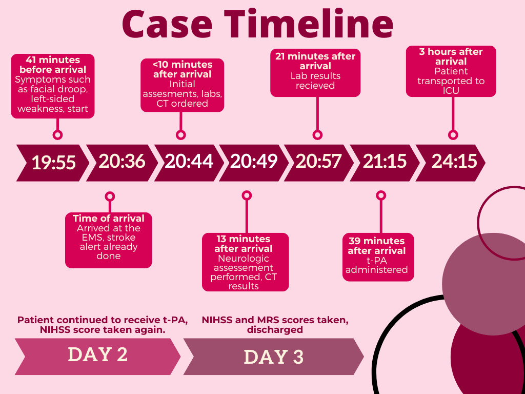
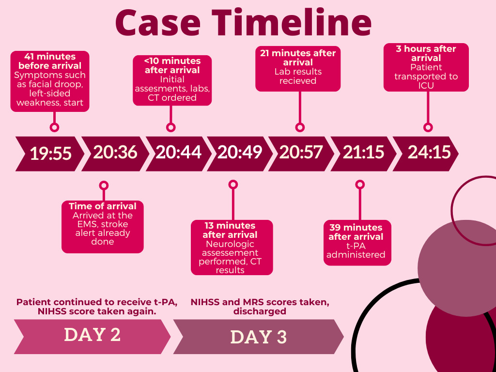
A 34-year-old Asian male presented to the emergency room with symptoms of a left-sided facial droop, slurred speech, and deep vein thrombosis after witnessing a generalized seizure at home. During the seizure, the patient did not fall due to a family member catching him. He first started exhibiting these symptoms 41 minutes prior to being brought in by emergency medical services as a stroke alert, which was called 10 minutes prior to the patient’s arrival (Figure 1).
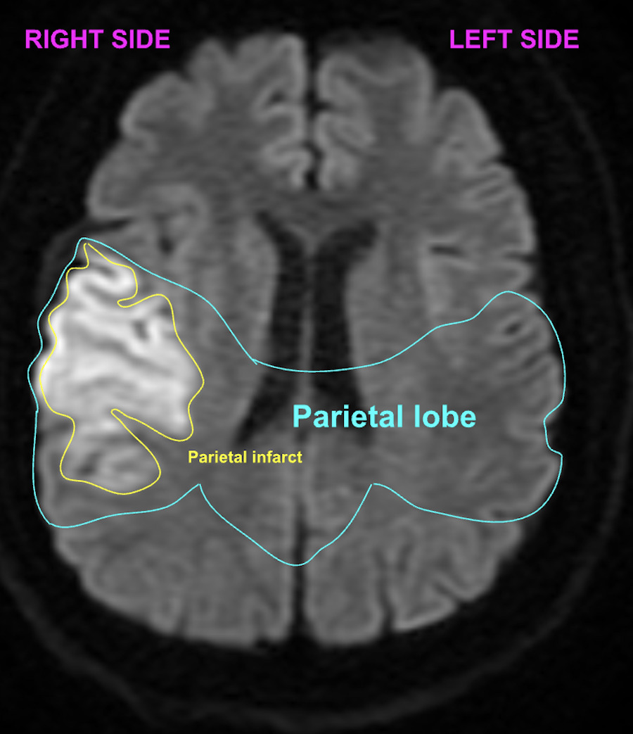
The patient had a medical history of atrial fibrillation and two prior cerebrovascular accidents that had not left any visible residual symptoms, due to which he was prescribed Plavix in the past. The patient’s vitals were stable upon arrival at the emergency room, and he was demonstrating a cardiac rhythm of atrial fibrillation. A neurologic examination revealed an initial National Institutes of Health Stroke Scale (NIHSS) score of 16 with left sided weakness and impaired speech. A brain computed tomography (CT) scan demonstrated no acute bleed or intracranial hemorrhage, and a computed tomography angiography (CTA) scan showed patent blood vessels. Through a magnetic resonance imaging (MRI) test, a moderately sized right parietal lobe infarction was seen (Figure 2).
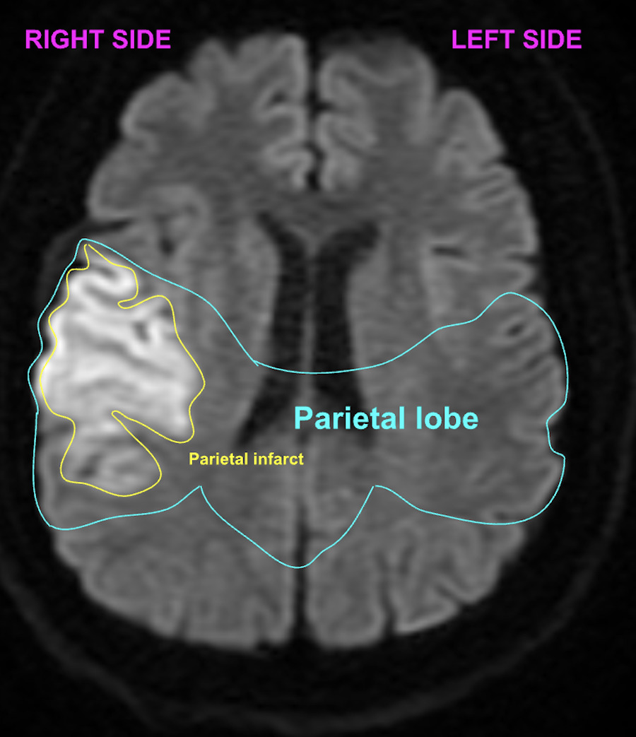
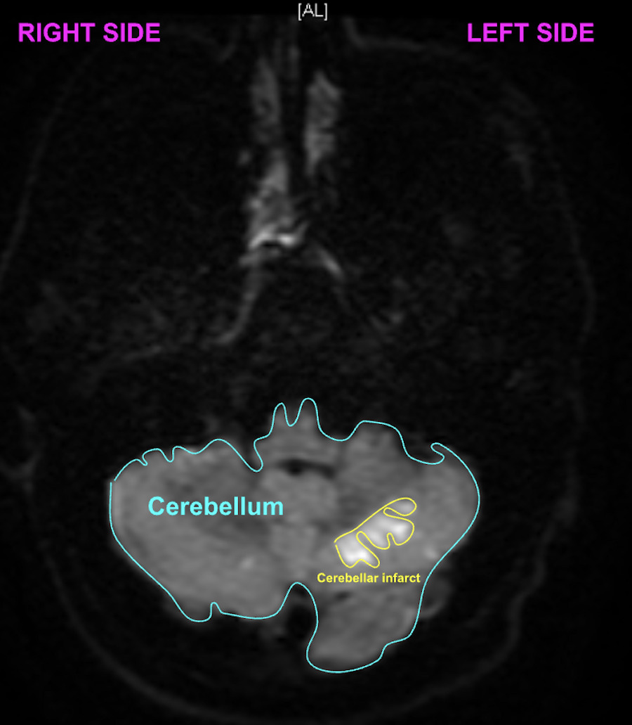
Multiple small, punctate infarctions in the left cerebellar hemisphere were found through another MRI scan in the posterior cranial fossa. Minimal infarction was observed in the right cerebellar hemisphere as well (Figure 3).
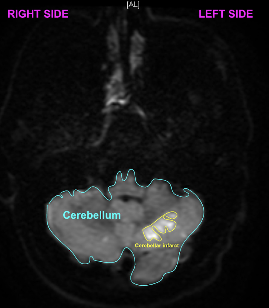
The patient was identified as a candidate for the thrombolytic agent tissue plasminogen activator (t-PA). The patient was thrombolysed 39 minutes after arrival and was admitted to the Intensive Care Unit (ICU) 3 hours after arrival. The patient’s NIHSS decreased to a score of 6 within one day of arrival, and to a score of 3 at the time of discharge. The Modified Rankin Scale (MRS) was performed, and the patient received a score of 1, still demonstrating low-intensity symptoms, but having no significant disability in performing basic, day-to-day tasks.
Discussion
This case demonstrates the diagnostic challenges of cerebellar infarctions and highlights the critical importance of rapid administration of tissue plasminogen activator (t-PA) in stroke management within an emergency department setting. Untreated cerebellar infarctions can progress to fatal cerebellar edema.13 Timely diagnosis and intervention in this case exemplifies the importance of prompt recognition and drug delivery in cerebellar strokes.
Cerebellar stroke diagnosis is challenging due to nonspecific symptoms, with dizziness occurring in 75% and nausea in over 50% of cases.14 Early symptoms often fail to suggest stroke, with more definitive signs like ataxia and paralysis emerging later. Accurate identification within three hours of symptom onset is essential for effective t-PA treatment and prevention of complications such as infarct-related edema. Imaging via MRI or CT can confirm cerebellar infarctions, but delays in initial clinical recognition often postpone these diagnostic measures. Misdiagnosis is common due to these challenges. Notably, the patient initially presented with standard stroke symptoms, including ipsilateral facial paralysis, dysarthria, and seizures, increasing a suspicion for stroke unlike most cerebellar stroke cases.12
Parietal infarctions, more common among ischemic strokes, further complicated this case by exacerbating the cerebellar stroke symptoms. This interaction expedited diagnosis and thrombolytic therapy. The patient’s history of atrial fibrillation and two prior strokes supported the clinical suspicion of vascular dysfunction. Ultimately, MRI confirmed infarctions in both the parietal and cerebellar regions.
Thrombolytic agent t-PA remains a critical ischemic stroke treatment, effective when administered within three to four and a half hours of symptom onset.15 Aspirin is commonly used for secondary prevention rather than acute treatment.16 In this case, the patient responded extremely well to t-PA, demonstrating its efficacy when administered promptly.
The patient’s history of recurrent strokes is noteworthy, particularly given the rarity of three ischemic strokes by the age of 34. Recurrence rates for ischemic strokes are approximately 9% and declining.17 However, atrial fibrillation increases recurrence risk by 54%.18 Despite being prescribed clopidogrel (Plavix) for secondary prevention, the patient experienced recurrent strokes.
Clopidogrel is approved for reducing recurrence risk after minor ischemic strokes or transient ischemic attacks (TIAs) during the first three months post-stroke. However, its long-term efficacy after a major ischemic stroke remains unclear. While clopidogrel prevents platelet aggregation in noncardioembolic strokes, evidence for its long-term benefit in recurrent stroke prevention is limited. Aspirin is favored for long-term secondary prevention, often in combination with clopidogrel per the American Heart Association guidelines. The lack of evidence supporting clopidogrel as a sole agent in acute ischemic stroke treatment raises concerns about its adequacy in high-risk patients as in this case.19,20
Further research is needed to assess the long-term efficacy of clopidogrel in recurrent stroke prevention. For patients with frequent vascular dysfunction, aspirin may be a more reliable alternative, especially when combined with other evidence-supported interventions. Addressing gaps in research is critical to optimizing treatment strategies and preventing recurrent strokes.
This case underscores the importance of emergency medicine physicians considering cerebellar stroke in patients presenting with atypical symptoms like dizziness and nausea, particularly in high-risk individuals with factors such as recurrent strokes, seizures, and atrial fibrillation. Prompt recognition is essential to prioritize the rapid administration of t-PA within the critical treatment window, reducing the risk of cerebellar edema and maximizing the chances of improved outcomes for patients.
Conclusion
The clinical presentation of cerebellar stroke can be affected by symptoms of additional infarctions in other neural structures. The use of thrombolytic agents is the widely used treatment for cerebellar and parietal ischemic stroke and have been proven to be more effective. This case stands out for the unconventional, clearer symptoms of cerebellar stroke that resulted in rapid diagnosis and treatment. It exemplifies how a timely diagnosis and immediate treatment can drastically improve ischemic stroke outcomes and highlights the effectiveness of t-PA as a clot-dissolving agent. Additionally, this case demonstrates how Clopidogrel may not be the best preventative measure for recurrence of ischemic stroke.
Consent to participate
Informed consent was obtained from the patient.
Consent to publish
The participant has consented to the submission of the case report to the journal.
Funding
No finding was received for this manuscript
Competing interests
None of the authors have any competing interests.
Ethics approval
The study was performed in accordance with the ethical standards as laid down in the 1964 Declaration of Helsinki and its later amendments or comparable ethical standards.
Data and/or Code availability
Not applicable
Authors’ contribution statement
All authors contributed to the case evaluation and write up. All authors agree to the final version of the manuscript.